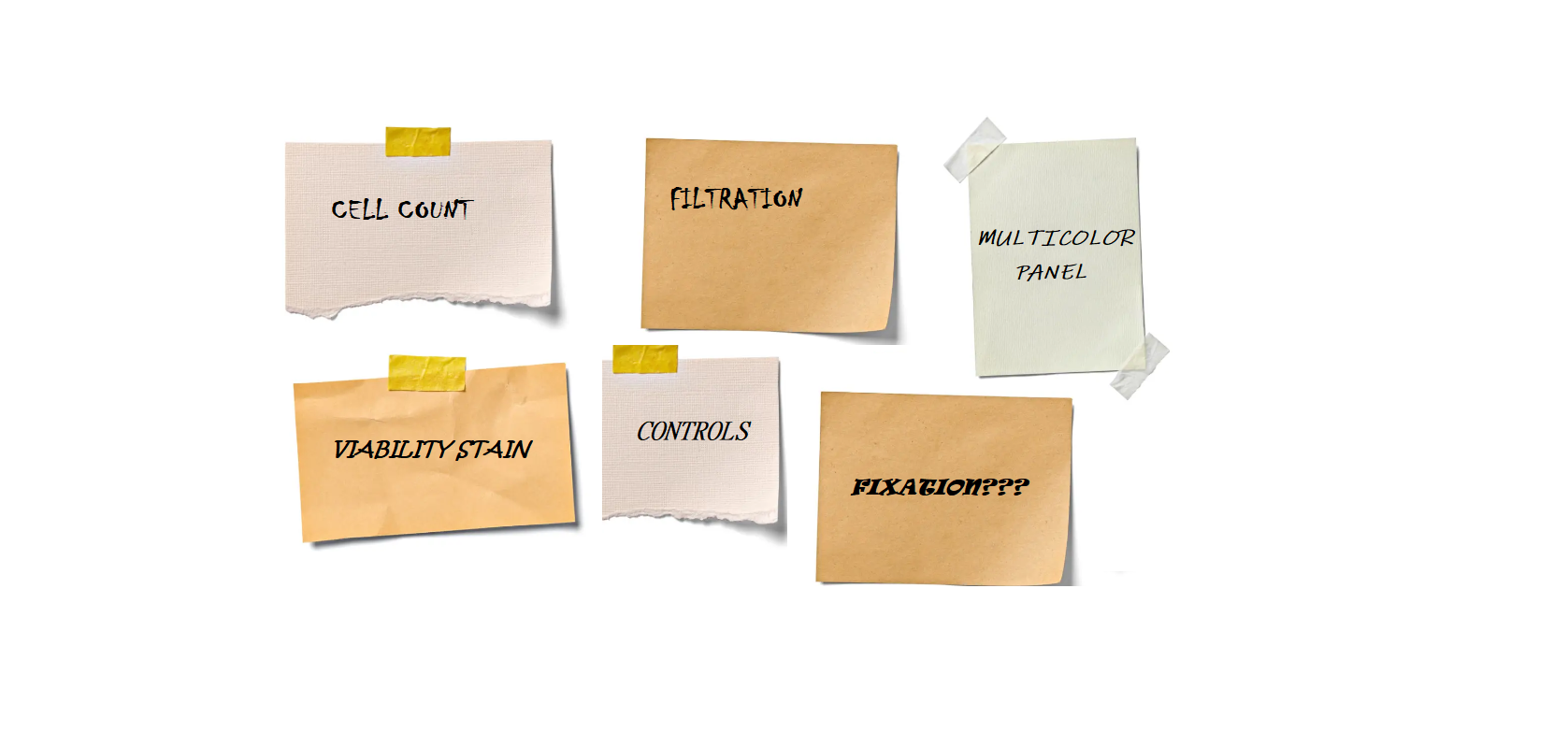
FLOW CITOMETRY TIPS
1. Count your cells
To ensure experimental uniformity, cells should always be counted prior to starting a flow cytometry experiment, regardless of the kind of material. For best results, a density of 105-107 cells/mL should be used; loading too few cells will lengthen workflows by increasing the time required to gather statistically significant numbers of events, while loading too many cells can result in events being lost from the analysis.
2. Eliminate the aggregates
The flow cytometer can become blocked by clumps of undesired debris, tissue fragments, and cellular aggregates. They ought to always be taken out before samples are acquired because they can also make data processing more difficult (doublets, for example, could be mistakenly recognized as dividing cells during cell cycle analysis). Cell strainers or DNase are two options available to you; the latter releases damaged cell DNA from buffers that may cause aggregation. Gating can be used to detect and remove the remaining aggregates from single-cell analyses.
3. Include a viability stain
Stains for cell viability offer an evaluation of the health of the cells and may warn researchers of any negative consequences of sample processing. Furthermore, they offer a more precise method than gating on light scatter qualities alone for removing dead cells and debris from cytofluorimetric analyses.
4. Extracellular marker staining: consideration on fixation
Even though both extracellular and intracellular markers are often measured in flow cytometry investigations, certain fixatives can modify the locations where antibodies attach to markers that are expressed on the cell surface. In order to avoid this, it is advised to stain extracellular markers before fixation; after that, cells can be permeabilized for the purpose of staining intracellular targets.
5. Include appropriate controls
Positive and negative controls are used to validate results and track the assay’s performance. Nevertheless, in order to guarantee accurate interpretation of fluorescent signals, flow cytometry needs the inclusion of multiple additional controls.
Unstained control: does not involve the use of antibodies. It guides the setting of background fluorescence.
Compensation controls: samples stained only with one fluorophore. Through them, spectral overlap can be measured.
Gating controls namely fluorescence minus one (FMO) controls: samples stained with all the fluorophores minus one fluorophore. They are used to measure fluorescence spread and better set true negative boundaries. You need these controls when you are analysing rare events, low expressed antigens or in any case you are working with samples that do not have a clear discrimination between negative and positive events.
6. Design the right multicolour panel
The relative amount of any target antigens and the characteristics of the fluorophores that will be utilized for detection are important factors to consider. It’s crucial to make sure that rare antigens and brilliant fluorophores are matched together as well as abundant antigens with dim fluorophores.
Chose fluorochromes to minimise spectral overlap. Spectral overlap can be avoided by choosing fluorochromes that are far apart or excited by different lasers. Sometimes spectral overlap cannot be avoided and signals have to be compensated. Compensation eliminates the non-specific signal generated by a fluorochrome in other detectors. This is why it is crucial to run single-label controls when setting up your experiment. Spectra viewers can hel you check if two or more fluorocromes can be used toghether. Have a look to BD® Spectrum Viewer.


