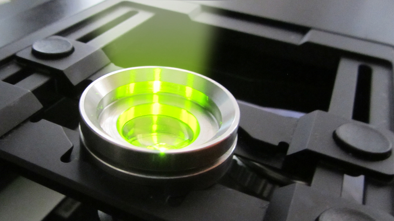
Advanced Microscopy and Imaging Core Facility
The facility houses a wide collection of instruments for imaging of not only biological samples.
It allows users to perform observation on a wide variety of methods, from basic observation with transmitted light to the more advanced fluorescent imaging technique on both living cells or tissues.
The facility offers highly specialized technical and scientific support throughout all the steps from the experimental design to the data analyses and presentation. A permanent senior technical expert is daily managing the instrument timetable and provides students training, research collaborations, commercial services to industry.
General information
Instructions are provided to users on the methodologies and the applications. The maintenance and operation of the equipment and the set-up of methods of analysis are coordinated by the staff in charge. Another task is the supervision of the planning, booking and report of devices. The analyses are carried out by the staff, in the presence of the users, but some routine analysis can be performed directly by selected users (expert-user) after specific training by a staff member (valid also for: Azure 400C, Histoline Cryostat, Histoline Microtome).
Applications:
- Transmitted and reflected light stereo microscopy.
- Transmitted light microscopy: bright-field, phase contrast.
- D.I.C. phase contrast for morphological live-imaging experiments
- Fluorescence wide-field microscopy.
- Laser scanning confocal microscopy.
- Time-lapse and in vivo imaging: bright-field, phase contrast, fluorescence wide-field, confocal laser scanning microscopy. With an incubation system to control environmental conditions.
- Advanced fluorescent techniques: FRET, FRAP, spectral acquisition, etc.
- Imaging of large samples such as mouse brain histological sections or drosophila larvae: mosaic, tiles and panorama modules.
- Molecular imaging, for protein gels or WB membranes: transmitted and reflected light, chemiluminescence, three channel fluorescence.
- Software for Image acquisition and analysis: ZEN Blue (Zeiss), NiS-Element F (Nikon), LAS AF(Leica), ImageJ-Fiji.
- Data analyses: intensity, colocalization, translocation, intensity profile, quantification, etc.
- Data presentation: image presentation, setup of graphical abstract, graphs for video presentation and paper figures.
Contact & info
Contact the staff before booking, asking about facility rates.
Facility Manager: Dr. Andrea Pagetta, PhD
Scientific Supervisor: Prof. Girolamo Calò
Staff: Dr. Andrea Pagetta, PhD; Dr. Alessia Forgiarini, PhD
Phone: 049.8275368/5082, email: andrea.pagetta@unipd.it
9:00 a.m. – 5:00 p.m. Monday-Friday
Location : DSF – Building C – L.go E. Meneghetti 2, Padua (Geotec 00200 -1 017)
Booking information
Ask the staff for booking. Time of slot 30 min. Booking, mandatory, here: <link>.
NOTE: Equipment are subject to usage charges.
NOTE: staff in charge must be contacted in very advance in case of experiments that will last more than three hours.
The staff provides assistance and support in teaching duties and to external users. The use of the instruments, supported by the staff, is also allowed to some “super-users”, experts in some specific fields, who can give their contribution to scientific research in terms of innovation in the development of new methods and new applications.


